Deconvolving Neuronal Ensembles from Macroscopic Neural Activities in Voltage-Sensitive Dye Image Streams on Mouse Cerebrum Slices
DOI:
https://doi.org/10.22452/mjs.vol45sp2.4Keywords:
Visual prosthesis, Neuronal ensemble, Voltage-sensitive dye imaging, Spatio-temporal cross-correlation analysesAbstract
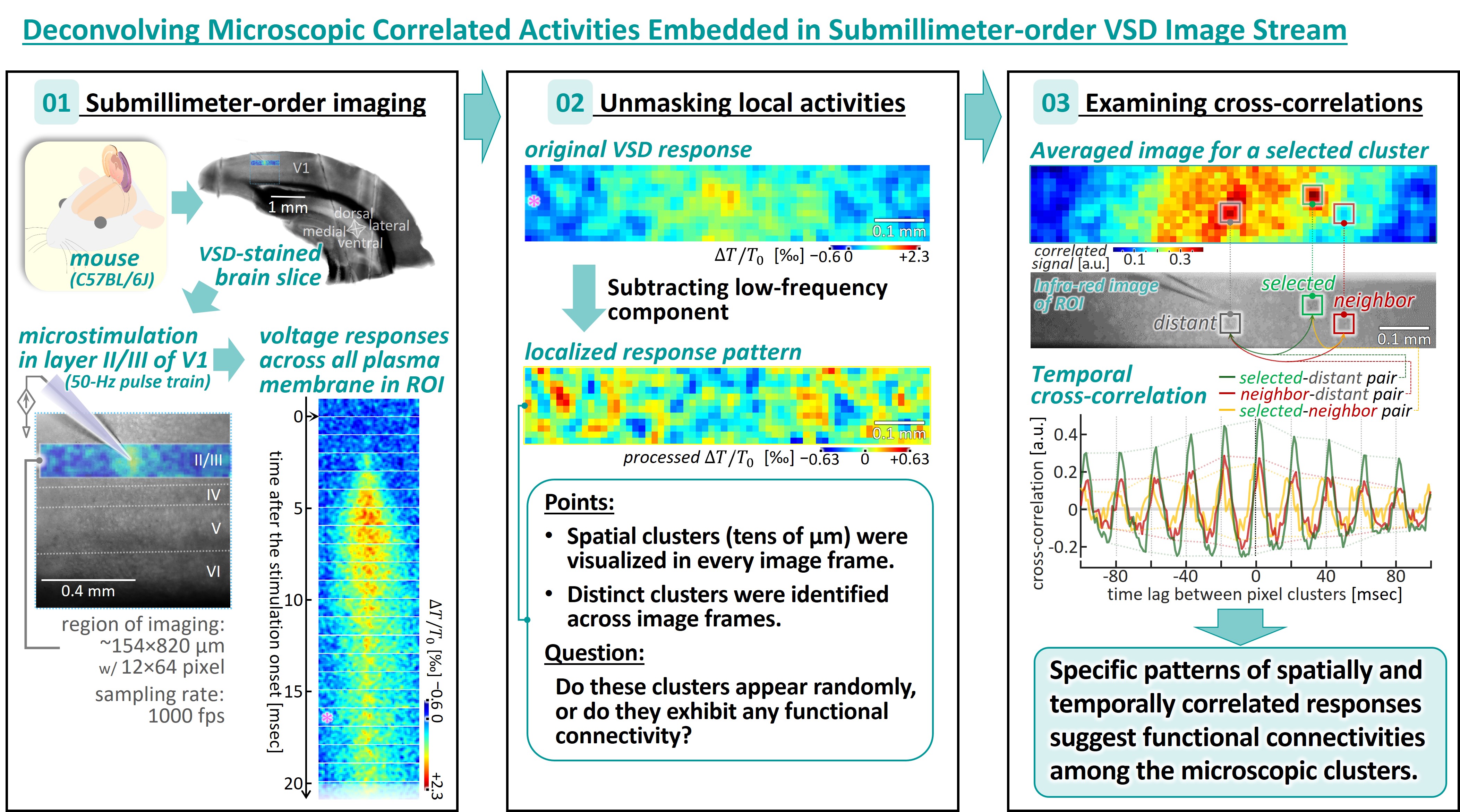
Previous clinical trials have demonstrated that electrical microstimulation delivered within the primary visual cortex (V1) in human subjects with acquired blindness can evoke focal light sensations in their visual fields, supporting the feasibility of intracortical visual prostheses. Our previous animal studies using voltage-sensitive dye (VSD) imaging examined the spatiotemporal patterns of cortical neural activity in response to electrical current-pulse stimuli, varying stimulus parameters to optimize stimulation design. These studies revealed specific relationships between stimulus parameters and neural responses at millisecond temporal resolution and submillimeter spatial scales. However, as VSD images typically represent voltage changes across all cytoplasmic membranes of cells responding to stimuli, activities of individual neuron populations and their functional connections are obscured by the macroscopic responses. This study aims to investigate stimulus-induced neuronal activities at a finer spatial resolution by employing image processing techniques. Specifically, spatial and temporal cross-correlation analyses were applied to VSD image streams to identify spatially distinguishable patterns of pixel clusters exhibiting temporally correlated VSD signals. In certain cases, these neuronal ensembles, each smaller than 100 micrometers in diameter, displayed time-fixed correlation in membrane voltage change, suggesting functional connectivity between them. The image processing method demonstrates the potential of separating distinct activities of cortical neuronal ensembles from macroscopic VSD imaging data, offering a valuable tool for detailed studies of stimulus-induced network dynamics and, in turn, optimizing stimuli for visual prostheses.
References
Benisty H., Song A., Mishne G., Charles A. S. (2022) Review of data processing of functional optical microscopy for neuroscience, Neuro-photonics, 9 (4): 041402 (32 pp.).
Chen X., Wang F., Fernández E., Roelfsema P. R. (2020) Shape perception via a high-channel-count neuroprosthesis in monkey visual cortex, Science 370 (6521): 1191-1196.
Fehérvári T. D., Okazaki Y., Sawai H., Yagi T. (2015) In vivo voltage-sensitive dye study of lateral spreading of cortical activity in mouse primary visual cortex induced by a current impulse, PLoS One 10 (7): e0133853 (24 pp.).
Fernández E., Alfaro A., Soto-Sánchez C., Gonzalez-Lopez P., Lozano A. M., Peña S., Grima M.D., Rodil A., Gómez B., Chen X., Roelfsema P. R., Rolston J. D., Davis T. S., Normann R. A. (2021) Visual percepts evoked with an intracortical 96-channel microelec-trode array inserted in human occipital cortex, J. Clin. Invest. 131 (23): e151331 (15 pp.).
Fukuda S., Nomoto T., Yoshida K., Hayashida Y., Yagi T. (2021) Accumulation of non-synaptic and synaptic excitations induced by microstimulation pulses in the mouse visual cortex layer II/III, 2021 43rd Ann. Int’l Conf. I.E.E.E. Med. Biol. Soc.: 5152.
Fukuda S., Yoshida K., Nomoto T., Yagi T., Hayashida Y. (2022) Use-dependent inactivation of the mouse visual cortex in response to microstimulation train, 2022 E-Health Bioeng. Conf.: 1-4.
Hayashida Y., Takeuchi K., Ishikawa N., Okazaki Y., Fehérvári T. D., Tanaka H., Yagi T. (2014) Voltage-sensitive dye imaging of the visual cortices responding to electrical pulses at different intervals in mice in vivo, 2014 36th Ann. Int’l Conf. I.E.E.E. Med. Biol. Soc.: 402-405.
Hayashida Y., Kameda S., Umehira Y., Ishikawa S., Yagi T. (2022) Multichannel stimulation module as a tool for animal studies on cortical neural pros-theses, Front. Med. Technol. 4: 927851 (21 pp.).
Miyamoto S., Suematsu N., Umehira Y., Hayashida Y., Yagi T. (2017) Age-related changes in the spatiotemporal responses to electrical stimulation in the visual cortex of rats with progressive vision loss, Sci. Rep. 7(1): 14165 (13 pp.).
Nomoto T., Tanaka Y., Hayashida Y., Yagi T. (2017) Imaging of population spikes induced by repetitive stimulus pulses in mouse cerebral slices in vitro, 2017 39th Ann. Int’l Conf. I.E.E.E. Med. Biol. Soc.: 1109.
Schmidt E. M., Bak M. J., F T Hambrecht, C V Kufta, D K O'Rourke, P Vallabhanath (1996) Feasibility of a visual prosthesis for the blind based on intracortical microstimulation of the visual cortex, Brain 119 (2): 507-522.
Tanaka Y., Nomoto T., Shiki T., Sakata Y., Shimada Y., Hayashida Y., Yagi T. (2019) Focal activation of neuronal circuits induced by microstimulation in the visual cortex, J. Neural Eng. 16 (3): 036007 (17 pp.).
Xie M. E., Adam Y., Fan L. Z., Böhm U. L., Kinsella I., Zhou D., Rozsa M., Singh A., Svoboda K., Paninski L., Cohen A. E. (2021) High-fidelity estimates of spikes and subthreshold waveforms from 1-photon voltage imaging in vivo, Cell Reports 35(1): 108954 (15 pp.).
Downloads
Published
Issue
Section
License
Copyright (c) 2026 Malaysian Journal of Science (MJS)

This work is licensed under a Creative Commons Attribution-NonCommercial 4.0 International License.
Transfer of Copyrights
- In the event of publication of the manuscript entitled [INSERT MANUSCRIPT TITLE AND REF NO.] in the Malaysian Journal of Science, I hereby transfer copyrights of the manuscript title, abstract and contents to the Malaysian Journal of Science and the Faculty of Science, University of Malaya (as the publisher) for the full legal term of copyright and any renewals thereof throughout the world in any format, and any media for communication.
Conditions of Publication
- I hereby state that this manuscript to be published is an original work, unpublished in any form prior and I have obtained the necessary permission for the reproduction (or am the owner) of any images, illustrations, tables, charts, figures, maps, photographs and other visual materials of whom the copyrights is owned by a third party.
- This manuscript contains no statements that are contradictory to the relevant local and international laws or that infringes on the rights of others.
- I agree to indemnify the Malaysian Journal of Science and the Faculty of Science, University of Malaya (as the publisher) in the event of any claims that arise in regards to the above conditions and assume full liability on the published manuscript.
Reviewer’s Responsibilities
- Reviewers must treat the manuscripts received for reviewing process as confidential. It must not be shown or discussed with others without the authorization from the editor of MJS.
- Reviewers assigned must not have conflicts of interest with respect to the original work, the authors of the article or the research funding.
- Reviewers should judge or evaluate the manuscripts objective as possible. The feedback from the reviewers should be express clearly with supporting arguments.
- If the assigned reviewer considers themselves not able to complete the review of the manuscript, they must communicate with the editor, so that the manuscript could be sent to another suitable reviewer.
Copyright: Rights of the Author(s)
- Effective 2007, it will become the policy of the Malaysian Journal of Science (published by the Faculty of Science, University of Malaya) to obtain copyrights of all manuscripts published. This is to facilitate:
- Protection against copyright infringement of the manuscript through copyright breaches or piracy.
- Timely handling of reproduction requests from authorized third parties that are addressed directly to the Faculty of Science, University of Malaya.
- As the author, you may publish the fore-mentioned manuscript, whole or any part thereof, provided acknowledgement regarding copyright notice and reference to first publication in the Malaysian Journal of Science and Faculty of Science, University of Malaya (as the publishers) are given. You may produce copies of your manuscript, whole or any part thereof, for teaching purposes or to be provided, on individual basis, to fellow researchers.
- You may include the fore-mentioned manuscript, whole or any part thereof, electronically on a secure network at your affiliated institution, provided acknowledgement regarding copyright notice and reference to first publication in the Malaysian Journal of Science and Faculty of Science, University of Malaya (as the publishers) are given.
- You may include the fore-mentioned manuscript, whole or any part thereof, on the World Wide Web, provided acknowledgement regarding copyright notice and reference to first publication in the Malaysian Journal of Science and Faculty of Science, University of Malaya (as the publishers) are given.
- In the event that your manuscript, whole or any part thereof, has been requested to be reproduced, for any purpose or in any form approved by the Malaysian Journal of Science and Faculty of Science, University of Malaya (as the publishers), you will be informed. It is requested that any changes to your contact details (especially e-mail addresses) are made known.
Copyright: Role and responsibility of the Author(s)
- In the event of the manuscript to be published in the Malaysian Journal of Science contains materials copyrighted to others prior, it is the responsibility of current author(s) to obtain written permission from the copyright owner or owners.
- This written permission should be submitted with the proof-copy of the manuscript to be published in the Malaysian Journal of Science
Licensing Policy
Malaysian Journal of Science is an open-access journal that follows the Creative Commons Attribution-Non-commercial 4.0 International License (CC BY-NC 4.0)
CC BY – NC 4.0: Under this licence, the reusers to distribute, remix, alter, and build upon the content in any media or format for non-commercial purposes only, as long as proper acknowledgement is given to the authors of the original work. Please take the time to read the whole licence agreement (https://creativecommons.org/licenses/by-nc/4.0/legalcode ).